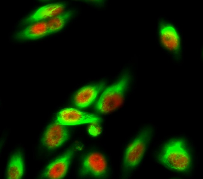
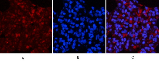
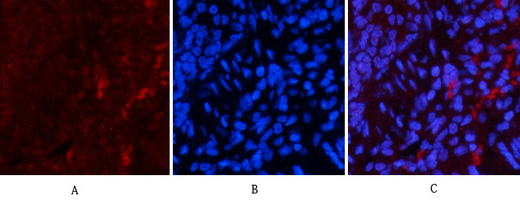
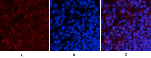
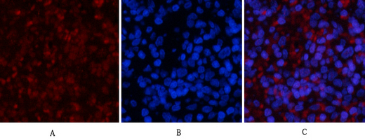
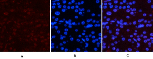
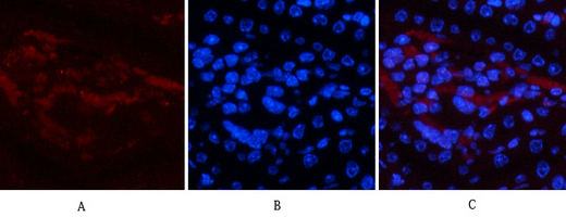
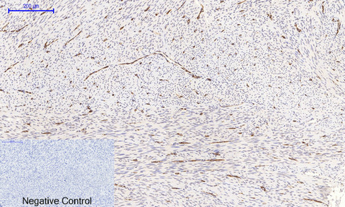
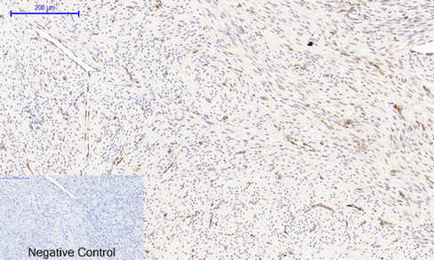
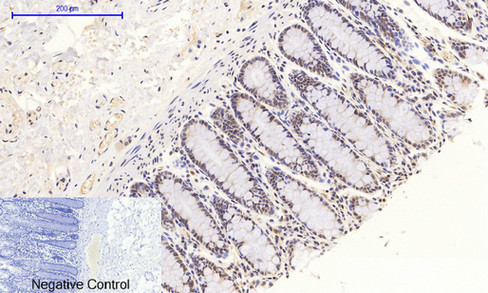
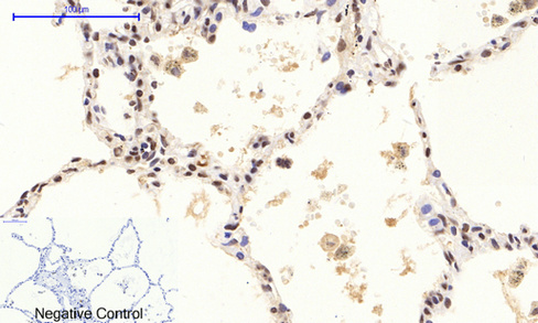
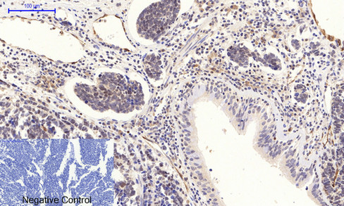
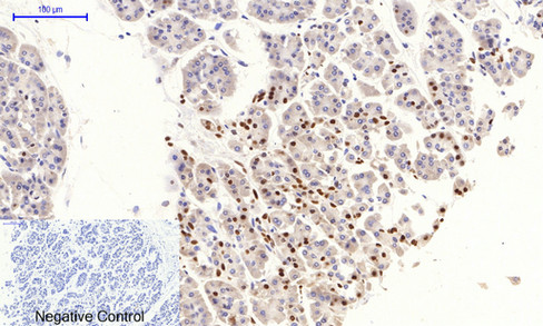
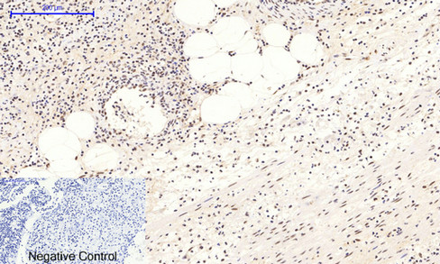
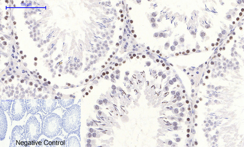
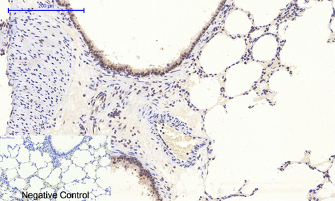
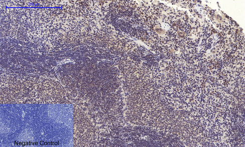
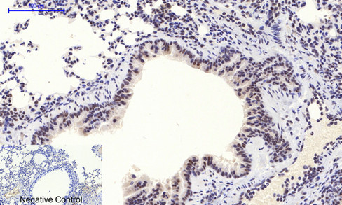
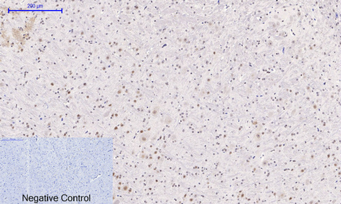
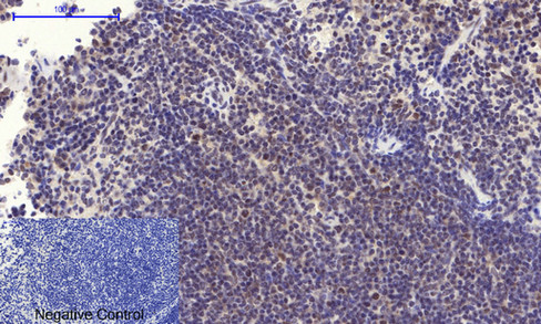
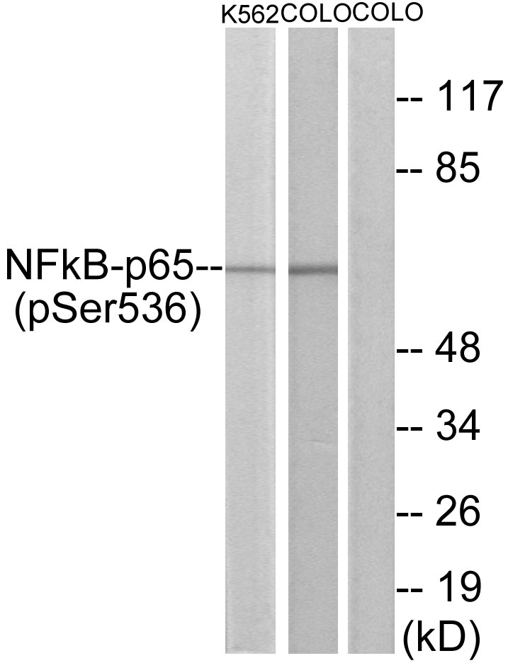
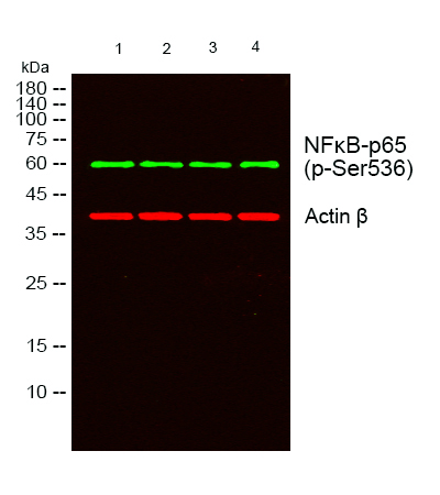
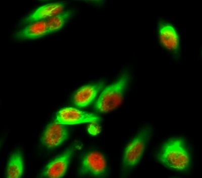
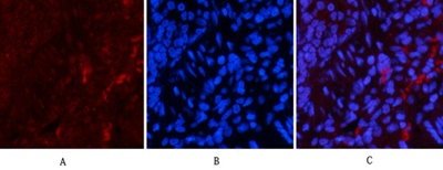
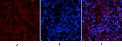
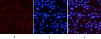
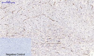
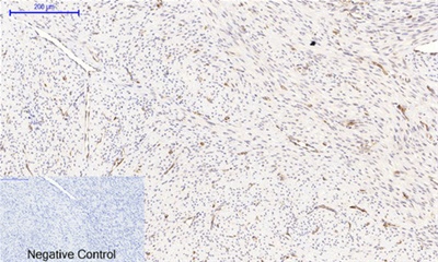
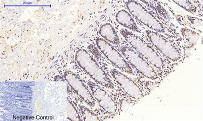
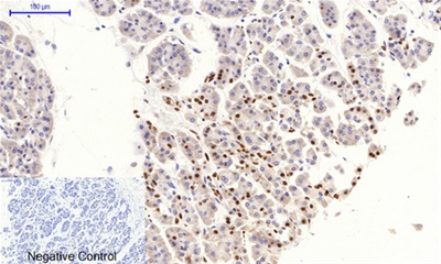
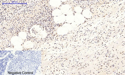
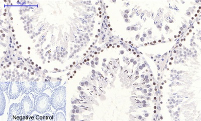
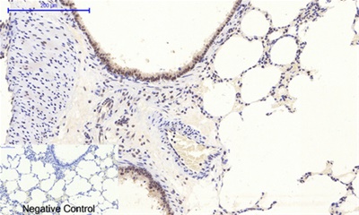
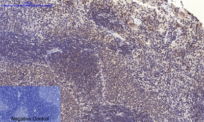
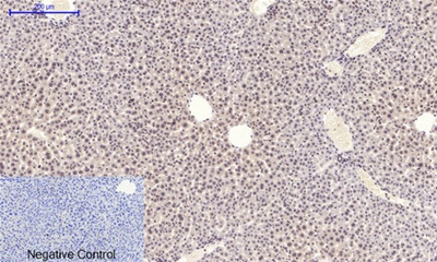
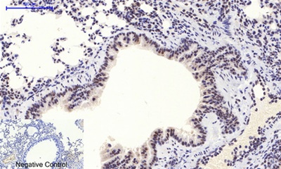
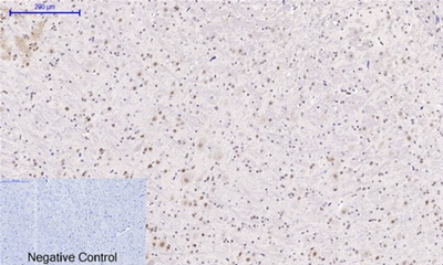
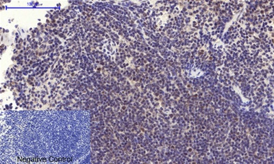
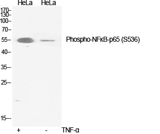
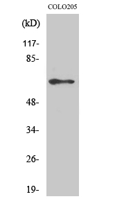
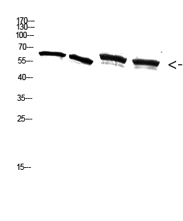
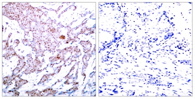
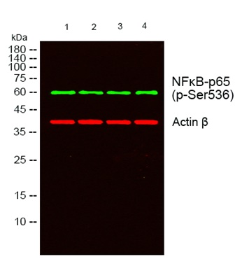
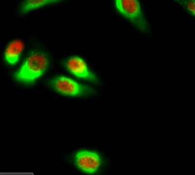
Immunofluorescence analysis of Hela cell. 1,NFκB-p65 (phospho Ser536) Polyclonal Antibody(red) was diluted at 1:200(4° overnight). α-tubulin Monoclonal Antibody(8F11)(green) was diluted at 1:200(4° overnight). 2, Goat Anti Rabbit Alexa Fluor 594 was diluted at 1:1000(room temperature, 50min). Goat Anti Mouse Alexa Fluor 488 was diluted at 1:1000(room temperature, 50min).Immunofluorescence analysis of rat-lung tissue. 1,NFκB-p65 (phospho Ser536) Polyclonal Antibody(red) was diluted at 1:200(4°C,overnight). 2, Cy3 labled Secondary antibody was diluted at 1:300(room temperature, 50min).3, Picture B: DAPI(blue) 10min. Picture A:Target. Picture B: DAPI. Picture C: merge of A+BImmunofluorescence analysis of rat-lung tissue. 1,NFκB-p65 (phospho Ser536) Polyclonal Antibody(red) was diluted at 1:200(4°C,overnight). 2, Cy3 labled Secondary antibody was diluted at 1:300(room temperature, 50min). 3, Picture B: DAPI(blue) 10min. Picture A:Target. Picture B: DAPI. Picture C: merge of A+BImmunofluorescence analysis of rat-spleen tissue. 1,NFκB-p65 (phospho Ser536) Polyclonal Antibody(red) was diluted at 1:200(4°C,overnight). 2, Cy3 labled Secondary antibody was diluted at 1:300(room temperature, 50min). 3, Picture B: DAPI(blue) 10min. Picture A:Target. Picture B: DAPI. Picture C: merge of A+BImmunofluorescence analysis of rat-spleen tissue. 1,NFκB-p65 (phospho Ser536) Polyclonal Antibody(red) was diluted at 1:200(4°C,overnight). 2, Cy3 labled Secondary antibody was diluted at 1:300(room temperature, 50min). 3, Picture B: DAPI(blue) 10min. Picture A:Target. Picture B: DAPI. Picture C: merge of A+BImmunofluorescence analysis of mouse-kidney tissue. 1,NFκBp65 (phospho Ser536) Polyclonal Antibody(red) was diluted at 1:200(4°C,overnight). 2, Cy3 labled Secondary antibody was diluted at 1:300(room temperature, 50min). 3, Picture B: DAPI(blue) 10min. Picture A:Target. Picture B: DAPI. Picture C: merge of A+BImmunofluorescence analysis of mouse-kidney tissue. 1,NFκBp65 (phospho Ser536) Polyclonal Antibody(red) was diluted at 1:200(4°C,overnight). 2, Cy3 labled Secondary antibody was diluted at 1:300(room temperature, 50min). 3, Picture B: DAPI(blue) 10min. Picture A:Target. Picture B: DAPI. Picture C: merge of A+BImmunohistochemical analysis of paraffin-embedded Humanuterus tissue. 1,NFκB-p65 (phospho Ser536) Polyclonal Antibody was diluted at 1:200(4°C,overnight). 2, Sodium citrate pH 6.0 was used for antibody retrieval(>98°C,20min). 3,Secondary antibody was diluted at 1:200(room tempeRature, 30min). Negative control was used by secondary antibody only.Immunohistochemical analysis of paraffin-embedded Humanuterus-cancer tissue. 1,NFκB-p65 (phospho Ser536) Polyclonal Antibody was diluted at 1:200(4°C,overnight). 2, Sodium citrate pH 6.0 was used for antibody retrieval(>98°C,20min). 3,Secondary antibody was diluted at 1:200(room tempeRature, 30min). Negative control was used by secondary antibody only.Immunohistochemical analysis of paraffin-embedded Humancolon tissue. 1,NFκB-p65 (phospho Ser536) Polyclonal Antibody was diluted at 1:200(4°C,overnight). 2, Sodium citrate pH 6.0 was used for antibody retrieval(>98°C,20min). 3,Secondary antibody was diluted at 1:200(room tempeRature, 30min). Negative control was used by secondary antibody only.Immunohistochemical analysis of paraffin-embedded Humanlung tissue. 1,NFκB-p65 (phospho Ser536) Polyclonal Antibody was diluted at 1:200(4°C,overnight). 2, Sodium citrate pH 6.0 was used for antibody retrieval(>98°C,20min). 3,Secondary antibody was diluted at 1:200(room tempeRature, 30min). Negative control was used by secondary antibody only.Immunohistochemical analysis of paraffin-embedded Humanlung-cancer tissue. 1,NFκB-p65 (phospho Ser536) Polyclonal Antibody was diluted at 1:200(4°C,overnight). 2, Sodium citrate pH 6.0 was used for antibody retrieval(>98°C,20min). 3,Secondary antibody was diluted at 1:200(room tempeRature, 30min). Negative control was used by secondary antibody only.Immunohistochemical analysis of paraffin-embedded Humanstomach-cancer tissue. 1,NFκB-p65 (phospho Ser536) Polyclonal Antibody was diluted at 1:200(4°C,overnight). 2, Sodium citrate pH 6.0 was used for antibody retrieval(>98°C,20min). 3,Secondary antibody was diluted at 1:200(room tempeRature, 30min). Negative control was used by secondary antibody only.Immunohistochemical analysis of paraffin-embedded HumanAppendix tissue. 1,NFκB-p65 (phospho Ser536) Polyclonal Antibody was diluted at 1:200(4°C,overnight). 2, Sodium citrate pH 6.0 was used for antibody retrieval(>98°C,20min). 3,Secondary antibody was diluted at 1:200(room tempeRature, 30min). Negative control was used by secondary antibody only.Immunohistochemical analysis of paraffin-embedded Rat-testis tissue. 1,NFκB-p65 (phospho Ser536) Polyclonal Antibody was diluted at 1:200(4°C,overnight). 2, Sodium citrate pH 6.0 was used for antibody retrieval(>98°C,20min). 3,Secondary antibody was diluted at 1:200(room tempeRature, 30min). Negative control was used by secondary antibody only.Immunohistochemical analysis of paraffin-embedded Rat-lung tissue. 1,NFκB-p65 (phospho Ser536) Polyclonal Antibody was diluted at 1:200(4°C,overnight). 2, Sodium citrate pH 6.0 was used for antibody retrieval(>98°C,20min). 3,Secondary antibody was diluted at 1:200(room tempeRature, 30min). Negative control was used by secondary antibody only.Immunohistochemical analysis of paraffin-embedded Rat-spleen tissue. 1,NFκB-p65 (phospho Ser536) Polyclonal Antibody was diluted at 1:200(4°C,overnight). 2, Sodium citrate pH 6.0 was used for antibody retrieval(>98°C,20min). 3,Secondary antibody was diluted at 1:200(room tempeRature, 30min). Negative control was used by secondary antibody only.Immunohistochemical analysis of paraffin-embedded Mouse-liver tissue. 1,NFκB-p65 (phospho Ser536) Polyclonal Antibody was diluted at 1:200(4°C,overnight). 2, Sodium citrate pH 6.0 was used for antibody retrieval(>98°C,20min). 3,Secondary antibody was diluted at 1:200(room tempeRature, 30min). Negative control was used by secondary antibody only.Immunohistochemical analysis of paraffin-embedded Mouse-lung tissue. 1,NFκB-p65 (phospho Ser536) Polyclonal Antibody was diluted at 1:200(4°C,overnight). 2, Sodium citrate pH 6.0 was used for antibody retrieval(>98°C,20min). 3,Secondary antibody was diluted at 1:200(room tempeRature, 30min). Negative control was used by secondary antibody only.Immunohistochemical analysis of paraffin-embedded Mousebrain tissue. 1,NFκB-p65 (phospho Ser536) Polyclonal Antibody was diluted at 1:200(4°C,overnight). 2, Sodium citrate pH 6.0 was used for antibody retrieval(>98°C,20min). 3,Secondary antibody was diluted at 1:200(room tempeRature, 30min). Negative control was used by secondary antibody only.Immunohistochemical analysis of paraffin-embedded Mousespleen tissue. 1,NFκB-p65 (phospho Ser536) Polyclonal Antibody was diluted at 1:200(4°C,overnight). 2, Sodium citrate pH 6.0 was used for antibody retrieval(>98°C,20min). 3,Secondary antibody was diluted at 1:200(room tempeRature, 30min). Negative control was used by secondary antibody only.Western Blot analysis of various cells using Phospho-NFκB-p65 (S536) Polyclonal Antibody diluted at 1:2000Western Blot analysis of COLO205 cells using Phospho-NFκBp65 (S536) Polyclonal Antibody diluted at 1:2000Western Blot analysis of A549 3T3 293T K562 cells using Antibody diluted at 2000. Secondary antibody was diluted at 1:20000Immunohistochemistry analysis of paraffin-embedded human breast carcinoma, using NF-kappaB p65 (Phospho-Ser536) Antibody. The picture on the right is blocked with the phospho peptide.Western blot analysis of lysates from K562 cells and COLO cells, using NF-kappaB p65 (Phospho-Ser536) Antibody. The lane on the right is blocked with the phospho peptideWestern blot analysis of lysates from 1) A549, 2) 3T3, 3) 293T , 4)K562 cells, (Green) primary antibody was diluted at 1:1000, 4°over night, secondary antibodywas diluted at 1:10000, 37° 1hour. (Red) Actin β Monoclonal Antibody(5B7) antibody was diluted at 1:5000 as loading control, 4° over night,secondary antibody was diluted at 1:10000, 37° 1hour.
RELA proto-oncogene, NF-kB subunit(RELA) Homo sapiens NF-kappa-B is a ubiquitous transcription factor involved in several biological processes. It is held in the cytoplasm in an inactive state by specific inhibitors. Upon degradation of the inhibitor, NF-kappa-B moves to the nucleus and activates transcription of specific genes. NF-kappa-B is composed of NFKB1 or NFKB2 bound to either REL, RELA, or RELB. The most abundant form of NF-kappa-B is NFKB1 complexed with the product of this gene, RELA. Four transcript variants encoding different isoforms have been found for this gene.
General Information
Reactivity
Human, Mouse, Rabbit, Monkey
Application
WB, IF, ELISA, IP, ICC, IHC-p
Host
Rabbit
Clonality
Polyclonal
Uniprot
Human Q04206;Mouse Q04207
Immunogen
The antiserum was produced against synthesized peptide derived from human NF-kappaB p65 around the phosphorylation site of Ser536. AA range:502-551


.png)