

General Information
| Reactivity | Mouse, Rat |
|---|---|
| Application | WB, IHC |
| Host | Rat |
| Clonality | Polyclonal |
| Conjugate | Non-conjugated |
| Alias | Macrosialin, Gp110, CD68 |
Figure :
|
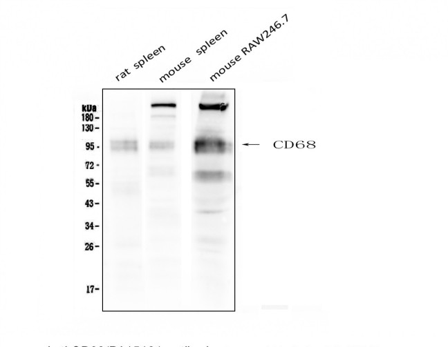
Western blot analysis of CD68 using anti- CD68 antibody. Lane 1: Rat Spleen Tissue Lysate Lane 2: Mouse Spleen Tissue Lysate Lane 3: Mouse RAW246.7 Tissue Lysate
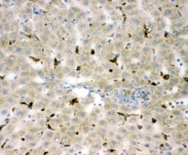
IHC analysis of CD68 using anti- CD68 antibody. CD68 was detected in paraffin-embedded section of Mouse Liver tissues. The tissue section was then incubated with 1μg/ml rabbit anti- CD68 Antibody overnight at 4°C. Biotinylated goat anti-rabbit IgG was used as secondary antibody and incubated for 30 minutes at 37°C. The tissue section was developed using Strepavidin-Biotin-Complex (SABC) with DAB as the chromogen.
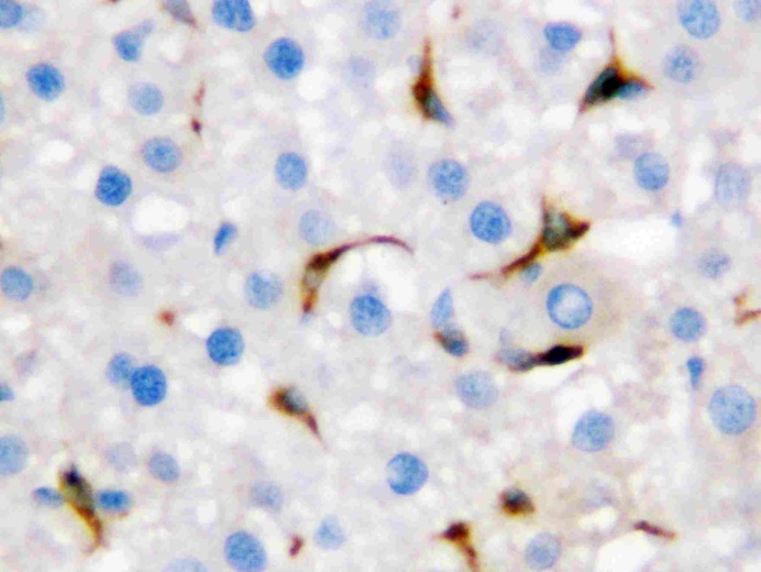
IHC analysis of CD68 using anti- CD68 antibody. CD68 was detected in paraffin-embedded section of rat liver tissues. The tissue section was then incubated with 1μg/ml rabbit anti- CD68 Antibody (PA1518) overnight at 4°C. Biotinylated goat anti-rabbit IgG was used as secondary antibody and incubated for 30 minutes at 37°C. The tissue section was developed using Strepavidin-Biotin-Complex (SABC) with DAB as the chromogen.
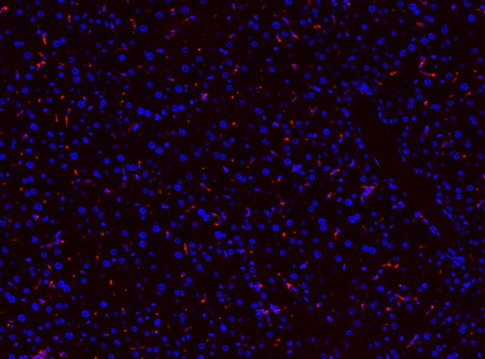
IF analysis of CD68 using anti- CD68 antibody. CD68 was detected in paraffin-embedded section of rat liver tissues. The tissue section was then incubated with 1μg/mL rabbit anti- CD68 Antibody overnight at 4°C. Cy3 Conjugated Goat Anti-Rabbit IgG was used as secondary antibody at 1:100 dilution and incubated for 30 minutes at 37°C. The section was counterstained with DAPI. Visualize using a fluorescence microscope and filter sets appropriate for the label used.
|
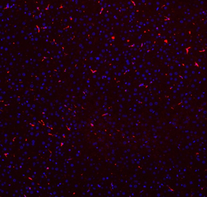
IF analysis of CD68 using anti- CD68 antibody. CD68 was detected in paraffin-embedded section of rat liver tissues. The tissue section was then incubated with 1μg/mL rabbit anti- CD68 Antibody overnight at 4°C. Cy3 Conjugated Goat Anti-Rabbit IgG was used as secondary antibody at 1:100 dilution and incubated for 30 minutes at 37°C. The section was counterstained with DAPI. Visualize using a fluorescence microscope and filter sets appropriate for the label used.


.png)