
General Information
| Reactivity | Human |
|---|---|
| Application | WB, IF, IHC, ELISA, IP |
| Host | Rabbit |
| Clonality | Polyclonal |
| Conjugate | Non-conjugation |
| Product Type | Primary Antibody |
|---|---|
|
Uniprot No. |
P55210 |
|
Immunogen |
Recombinant Human Caspase-7 protein (24-198AA) |
| Application |
WB : 1:1000-5000 IHC : 1:200-500 IF : 1:50-200 IP : 1:200-2000 |
|---|
| Figure |
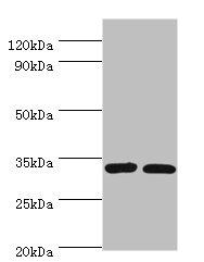
Figure 1 : Western blot analysis using Caspase-7 antibody at 2µg/ml
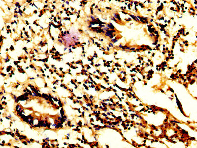
Figure 2 : IHC image with A8926P diluted at 1:200 and staining in paraffin-embedded human appendix tissue performed on a Leica BondTM system. After dewaxing and hydration, antigen retrieval was mediated by high pressure in a citrate buffer (pH 6.0). Section was blocked with 10% normal goat serum 30min at RT. Then primary antibody (1% BSA) was incubated at 4°C overnight. The primary is detected by a biotinylated secondary antibody and visualized tissue using an HRP conjugated SP system.
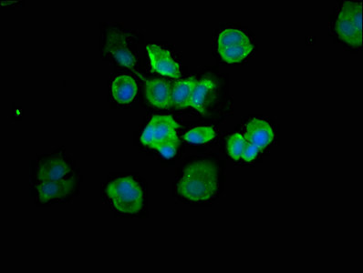
Figure 3 : Immunofluorescence staining of MCF-7 cells with A8926P at 1:66, counter-stained with DAPI. The cells were fixed in 4% formaldehyde, permeabilized tissue using 0.2% Triton X-100 and blocked in 10% normal Goat Serum. The cells were then incubated with the antibody overnight at 4°C. The secondary antibody was Alexa Fluor 488-congugated AffiniPure Goat Anti-Rabbit IgG (H+L). |
|---|---|
| Purification Method | >95%, Protein G purified |
| Storage Buffer | Preservative: 0.03% Proclin 300 Constituents: 50% Glycerol, 0.01M PBS, PH 7.4 |
| Storage Instruction | Upon receipt, store at -20°C or -80°C. Avoid repeated freeze. |
|---|
| Alias | Caspase-7 (CASP-7) (EC 3.4.22.60) (Apoptotic protease Mch-3) (CMH-1) (ICE-like apoptotic protease 3) (ICE-LAP3) [Cleaved into: Caspase-7 subunit p20; Caspase-7 subunit p11], CASP7, MCH3 |
|---|


.png)