


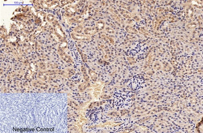
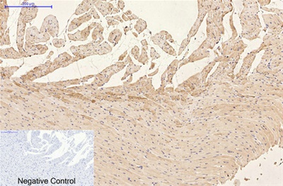
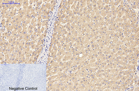
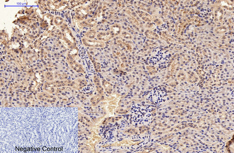
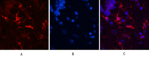
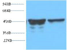
GFAP Monoclonal Antibody(5C8)
- Catalog Number : A20465PI
- Number :
-
Size:
Qty : - Price : Request Inquiry
-


Introduction
glial fibrillary acidic protein(GFAP) Homo sapiens This gene encodes one of the major intermediate filament proteins of mature astrocytes. It is used as a marker to distinguish astrocytes from other glial cells during development. Mutations in this gene cause Alexander disease, a rare disorder of astrocytes in the central nervous system. Alternative splicing results in multiple transcript variants encoding distinct isoforms. [provided by Ref Seq, Oct 2008],
General Information
| Reactivity | Human, Mouse, Rabbit |
|---|---|
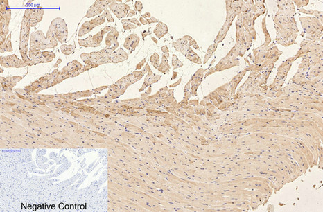
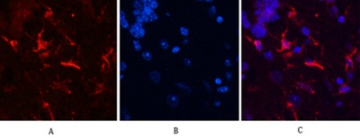
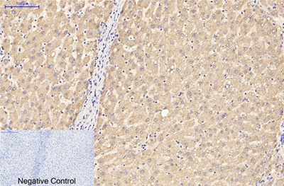
| Application | WB, IF, IHC |
| Host | Mouse |
| Clonality | Monoclonal |
| Conjugate | Non-conjugation |
| Uniprot | Human: P14136 / Mouse: P03995/ Rat: P47819 |
| Immunogen | Synthetic Peptide of GFAP |
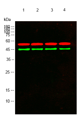
| Assay principle | WB:1:2000-1:5000/IHC:1:50-1:300/IF:1:200 |
| Purity | The antibody was affinity-purified from mouse ascites by affinity-chromatography using specific immunogen. |
| Formula | PBS, pH 7.4, containing 0.5%BSA, 0.02% sodium azide as Preservative and 50% Glycerol. |
| Storage instruction | Store at -20℃ for 1 year. |
| Alias | GFAP; Glial fibrillary acidic protein; GFAP |
|
|


.png)